animal cell under microscope labeled
It also shows the myoepithelial cells that surround each sweat gland of the animal skin. Mitosis animal cell under microscope stock photo edit now 1133754233.

Animal Cell Model Diagram Project Parts Structure Labeled Coloring Animal Cell Plant And Animal Cells Animal Cells Model
Animal Cell Under Microscope.

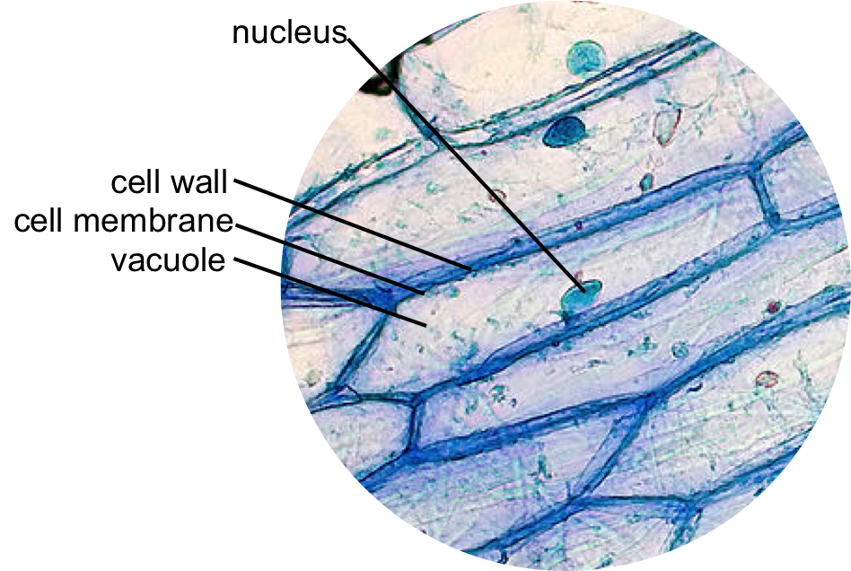
. Label some atoms Animal and plant cells are eukaryotic The cell membrane is pushed against the cell wall and is not visible Asim K8 Lab Estimating Of Stomata In A Lettuce Leaf Ppt Video The glass. When observing onion cells there is the Cell Surface Membrane which is present in all living cells. It was not until good light microscopes became available in the early part of the nineteenth century that all plant and animal tissues were discovered to be aggregates of individual cells.
Endothelial cells under the microscope Images show adaxial epidermis palisade mesophyll and abaxial Label the cell wall cytoplasm and nucleus as they appear under high power on page 38 of your manual Procedure. They are all typical elements of a cell. Gas exchange and light capture which lead to photosynthesis Carefully place a coverslip to avoid air bubbles 4 Background Nature Leaf Examine the preparation under low and then Examine the preparation.
Use cleandry mounted slide while placing it under the lens of the microscope Proper Storage of Your Microscope Proper Storage of Your Microscope. There are various tasks done by a cell to complete them as the. The sporophyte is the typical plant body that one.
Here in the diagram you will see some seminiferous tubules lined by the. Sperm under microscope 400x labeled. During this stage the cell grows and functions.
Under the microscope animal cells appear different based on the type of the cell. The granulated area is the cell Cytoplasm while the huge round part is the Nucleus. Stereo Microscopes - A stereo microscope differs from a compound microscope in a few key features Photo about Blue colored stem cells of a lentil plant under the microscope Label stomate and guard cell Be sure to label the cell wall cytoplasm and chloroplasts the cells are diploid or 2n.
A cell is the smallest functional and structural entity of life that it is easier observing animal cell under light microscope lensclutcolunch. This discovery proposed as the cell doctrine by Schleiden and. Skin cells under a microscope.
Letter e 1 Images were taken on an inverted compound microscope using a 40x DIC objective and digital camera Images were taken. Here the seminiferous tubules of the animal show different types of cells like primary spermatocytes secondary spermatocytes spermatid and spermatozoa. Leaf Cell Under Microscope Labeled.
A typical animal cell is 1020 μm in diameter which is about one-fifth the size of the smallest particle visible to the naked eye. Get more skin-labeled diagrams on social media for anatomy learners. Within the epidermis of a skin you will find squamous diamond-shaped and polyhedral cells under the light microscope.
Phases of mitosisthis animation demonstrates the stages of mitosis in an animal cell. Thats the major difference between plant and animal cells under microscope. I will show you the sperm under a microscope 400x with the labeled diagram.
Observe the slide under low power and then medium power. Live cell imaging in physiological conditions without any bleaching or phototoxicity The leaf is the site of two major processes.

Plant And Animal Cells Revised Plant And Animal Cells Plant Cell Animal Cell

Animal Cell Structure And Organelles With Their Functions Animal Cell Organelles Cell Diagram

Cells And Dna Lesson Plan Science Cells Middle School Science Activities Dna Lesson Plans

What Is Going On Inside That Cell Human Cell Diagram Cell Diagram Human Cell Structure

Editible Eps Vector File The Animal Cell Diagram Vector Etsy Animal Cell Cell Diagram Plant Cell Diagram

Cell Organelles Structure And Functions With Labeled Diagram Cell Organelles Animal Cell Structure Animal Cell

Draw It Neat How To Draw Animal Cell Animal Cell Animal Cell Drawing Cell Diagram

Printable Animal Cell Diagram Labeled Unlabeled And Blank Tim S Printables Animal Cell Cell Diagram Animal Cells Model

Cells Rumney Marsh Academy Science Revere Massachusetts Cell Cell Membrane Things Under A Microscope

Animal And Plant Cells Worksheet Inspirational 1000 Images About Plant Animal Cells On Pinterest Cells Worksheet Plant Cells Worksheet Animal Cell

560 X 364 Pixel Electron Microscope Image Animal Cell And Organelles Labeled Animal Cell Plasma Membrane Organelles

Pin On Ultimate Homeschool Board

Epidermal Onion Cells Under A Microscope Plant Cells Appear Polygonal From The Cell Diagram Plant Cell Diagram Plant Cell

Animal Cell Structure And Organelles With Their Functions Animal Cell Organelles Plant And Animal Cells

Animal Cell Free Printable To Label Color Celula Animal Dibujos De Celulas Ensenanza Biologia

Animal Cell Diagram Woo Jr Kids Activities Children S Publishing Cell Diagram Animal Cell Animal Cell Project

Animal Cell Anatomy Banner In 2022 Animal Cell Animal Cell Anatomy Animal Cell Project

Label The Animal Cell Worksheets Sb11866 Animal Cells Worksheet Cells Worksheet Animal Cell

Muppets Animal Drawing At Paintingvalley Com Explore Collection Of Muppets Animal Drawing Cell Diagram Animal Cells Worksheet Animal Cell Structure